


Imaging consists of different types of microscopy (optic-, fluorescence-, confocal and electron microscopy), that allow in vitro, in vivo and ex-vivo studies of samples at high magnification, aimed at characterising subcellular structures and organisms. In vivo imaging allows to reduce the number of experimental animals by longitudinal studies, in which the same animal is used at different time points. Moreover, in vivo imaging decreases animal suffering since procedures are usually performed under deep anaesthesia.
- LEICA TCS-SP5 Inverted Confocal Microscope. This microscope has an inverted stage, equipped with different objectives (dry 5-10-20x , high NA oil immersion 40 e 63x, glycerol 63x, oil 100x; different Phase contrast objectives are also available). The microscope is equipped with 4 visible lasers emitting the following wavelengths: 405nm, 458nm, 476nm, 488nm, 496nm, 514nm, 543nm, 633nm. The signal can be detected by 5 spectral detectors (2 Hybrid and 3 PMTs). An external PMT is available for the bright field signal. A motorised stage allows mosaic acquisition and stitching (Tile Scan), as well as time lapse on different points of the sample at the same time (Mark and Find). The incubator, equipped with temperature and CO2 controller, allows to perform live cell imaging for long hours.
The software is equipped with FRET-AB, FRET-SE, FRAP, live data mode wizards.
A 8 kHz resonant resonant scanner is also available to reduce bleaching during live acquisition.
Recently, this machine has been equipped with a widefield acquisition system including a high speed fluorescence camera (Hamamatzu), Metamorph acquisition software and the Gemini system for widefield FRET. -
LEICA MYCROSYSTEMS- STELLARIS WLL DIVE FALCON Confocal-Multiphoton Microscope: an upright microscope that can be operated in confocal, multiphoton, and FLIM modes. The system is equipped with an HCX-PL-APO NA 1.0 objective for multiphoton microscopy, as well as long working distance objectives (HCX APO L 10x/0.30 W, HCX APO L 20x/0.50 W U-V-I -/D 3.5, HCX APO L 40x/0.80 W UVI, HCX APO L 63x/0.90). The microscope stand features a motorized stage for mosaic imaging, navigation, and multipoint acquisition. The system is equipped with a confocal scan head including two internal detectors (HyDX and HyDS) and a pulsed white-light laser covering the 440–680 nm wavelength range. The detectors can operate either in standard mode or in semi-quantitative FLIM (Tau) mode. The multiphoton module is equipped with a Chameleon Discovery NX laser (Coherent) providing two excitation wavelengths: one fixed at 1040 nm and one tunable from 690 to 1300 nm. It also includes the DIVE module with two non-descanned detectors (NDDs), consisting of one HyDX detector and one PMT detector. The FALCON module enables FLIM operation in both confocal and multiphoton modes, allowing improved separation of spectrally overlapping fluorophores, removal of autofluorescence, and other applications based on fluorescence lifetime measurements. Multiphoton microscopy is a fluorescence optical microscopy technique in which the sample is excited at the focal point by pulsed infrared laser light. The use of long wavelengths provides: (i) greater penetration depth of the incident radiation compared with visible light, and (ii) minimal fluorescence photobleaching. A typical application of multiphoton microscopy is the observation of eukaryotic cells in thick specimens such as organoids, lymph nodes, tissue slices, or in vivo animal models. Examples include imaging neurons or microglia in the cerebral cortex of transgenic mice expressing fluorescent proteins.
- EVIDENT FV4000 Inverted confocal microscope, equipped with 4-10-20x air, long distance 20X air, 30x Silicon e 60x oil immersion objectives). 8 Laser Lines (405, 445, 488, 514, 561, 594, 640, 785 nm). Detection: up to 6 SiPM 16 bit detectors operating in photon counting. Extended detection up to 900nm. Bright field detector available. The motorized stage allow to make mosaic and multi area time lapse. The system is equipped with a cage incubator for temperature, CO2 and humidity control for live imaging experiments. The system is equipped with resonant scanner 1024×1024, focus control, integrated deconvolution, Cell Sens software for analysis.
- Workstation for image analysis with the following software: IMARIS 10.2 (Andor), CellSens (Evident), Huygens (SVI).
- Optical Imager IVIS Lumina S5 Revvity. The system allows to make a fluorescence or bioluminescence in vivo and ex-vivo imaging. It is equipped with a -90°C chilled Charge Coupled Device (CCD) with quantum efficiency greater than 85% between 500 and 700 nm and greater than 30% between 400 and 800 nm. The system is equipped with a broad range of excitation and emission filters in order to allow a complete acquisition in the fluorescence mode, allowing spectral unmixing. The system is also provided with a license for multimodal imaging in order to combine images from different imaging modalities (TC, PET, Magnetic Resonance and OI) The Optical Imager technique is used to detect optical photons, especially in the red and infrared regions. The most relevant applications are the fluorescence ( excitation of fluorophore injected in organism) and bioluminescence (enzymatic reactions creating light) detection. The OI technique is very sensitive (at the level of single cells), reliable, fast and cheap.
In the fluorescence mode, the instrument uses the fluorescence emission of colorant injected in living organism. Moreover, different kinds of cells (including stem cells) can be marked with fluorescent molecules allowing to study their homing in vivo. In the bioluminescence mode, OI detects light emitted in specific enzymatic reactions. Here in Verona, the Cerenkov Luminescence Imaging (CLI) has been discovered and developed, a new technique combining Optical Imaging and Nuclear Medicine. The CLI technique has been already used in humans. - Magnetic Resonance Imaging Tomograph (Bruker, Biospin) for small animals. MRI system is equipped with a 7 Tesla, 16 cm bore Ultra Shilded superconducting magnet (Pharmascan 70/16 US Bruker). It’is based on Bruker Advance II electronics and a Bruker B-GA9S HP gradient insertwith 380 mT/m maxim intensity. It’s equipped with five acquisition coils: birdcage coil for rats and mice, helmet surface coil for rats and mice brain and array coil- 4 channels for mouse brain. For in vivo acquisition, the system is provided with gas anesthesia, vital signs measurement and heating devices for animals. In oncological research, MR standard images are used to study tumor growth and to distinguish tumor tissue from perifocal edema. At the same time, in the Central Nervous System, MR standard images allow to evaluate lesions, blood brain permeability and atrophy. Advanced MRI application allow to measure blood flow (using the Artery Spin Labeling technique), to evaluate the functional answer to a stimulus (with the BOLD technique), the axonal connectivity (with the Diffusor Tensor Imaging technique) and the functional connectivity (resting state functional MRI). Finally, at high field, localized Magnetic Resonance Spectroscopy (MRS) allows not invasive detection and quatification of different metabolites that have crucial relevance for staging tumors and defining CNS pathologies, like N-Acetil Aspartate (NAA), choline (Cho), creatine (Cr, myo inositol (MI) and glutamate and glutamine compounds (Glu-n).
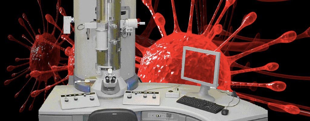
- Transmission Electron Microscope JEOL-JEM2100-Plus (TEM). A high-resolution imaging system equipped with an integrated digital camera. The TEM provides solutions for a wide range of challenges in materials science, nanoelectronics, and biological sciences. It enables two-dimensional observation of biological or inorganic samples with extremely high resolution (2-3 nm). The TEM, featuring a LaB6 thermionic electron source, is equipped with accessories such as STEM (HAADF and BF) and EDS for microanalysis. The maximum magnification achievable with this instrument is 1,000,000×.
For info and booking, see the Access and Booking page.